Case Report
Anti-Gravitational Growth-Associated Changes in the Mobilization of Leg Muscles during Walking in One Girl
Luna Ohira1,*, Takashi Ohira2,*, Koji Kitada3, Tomotaka Ohira4, Fuminori Kawano5, Taku Wakahara6, Kiyotaka Kamibayashi6, Yasuo Nakamura6, Yoshinobu Ohira6,7
1Faculty of Health and Well-being, Kansai University
2Division of Aerospace Medicine, Department of Cell Physiology, The Jikei University School of Medicine
3Ishikawa National College of Technology
4Chiben Gakuen, Nara College
5Graduate School of Health Sciences, Matsumoto University
6Graduate School of Health and Sports Science, Doshisha University
7Research Center for Adipocyte and Muscle Science, Doshisha University
*:Equally contributed authors
ABSTRACT
Growth-related changes in the mobilization of leg muscles during walking were studied in human subject. Mobilization of soleus, lateral portion of gastrocnemius (LG), and tibialis anterior (TA) muscles during the first walking steps in the life of a 12-month and 11-day old baby girl was checked by recording electromyogram (EMG). The same recordings were also performed, when she became 15 years old. The body balance during walking was also monitored by recording the ground reaction forces on a three-dimensional force plate. Co-contraction of ankle plantar-flexors (soleus and LG) and dorsi-flexor (TA) was observed during the first walking in her life. The EMG amplitudes and the step intervals were not constant. But disturbance of body balance toward both anterior/posterior and right/left directions was minor, although some degree of fluctuation was noted in the vertical force during the body-support phase. The relatively stable forces toward anterior and posterior direction, without heel-strike followed by toe-kick, suggested that her landing patterns were flat-foot strike. Data also suggested that the immature walking was not directly related to the poor body balance. The co-contraction of antagonists disappeared and walking patterns were normalized at the age of 15. It was speculated that afferent input may play an important role in the learning of walking motor performance in new-born babies, since the afferent neural input is closely related to the antigravity activity of muscle.
Abbreviations: LG, lateral portion of gastrocnemius; TA, tibialis anterior; EMG, electromyogram
(Received:9 May, 2015 Accepted:29 June, 2015)
Key words:First independent step of an infant, mobilization of lower leg muscles, body balance
INTRODUCTION
It has been well-known that children improve their upright gait patterns from new-born stepping to mature walking following the growth and development1-3,5,6,19-23). Supported walking patterns and mobilization of leg muscles were analyzed in new-born baby as early as 3 weeks20) or 1 month23) after birth. Co-contraction of mutual antagonists was the typical phenomena noted in neonates during walking3,19,20,23). However, such mobilization of muscles was changed to reciprocal recruitment of antagonists following the growth. Further, it was reported that transition of gait patterns was induced from digitigrade to plantigrade1) or from initial contact with toes, foot-flat or heel to consistent heel strike21) during the first year after independent walking. It is well-known that the antagonistic dorsi- and plantar-flexors are recruited alternately in each step during walking of matured human15) and animals18).
It was speculated that such growth-related changes might be related to the increased muscle strength19,20) and/or improved balance due to neuro-maturation1,19,20). However, it is not clear how infants, who are not experienced the independent bipedal walking in gravitational environment, utilize their lower leg muscles during walking without any support. Therefore, the current study was performed to investigate the mobilization of dorsi- and plantar-flexors during the first independent walking in the life and after 15-year-growth in human subject. Further, the body balance during walking was also checked recording the three-dimensional ground reaction forces using a force plate.
MATERIALS AND METHODS
This study was performed using one female subject. The parents of a baby girl and herself at 15-year old were informed about the possible risks in attending the walking exercise and recording of electromyogram (EMG). And signed informed consents were obtained from them. This study was approved by the Human Use Committee at National Institute of Fitness and Sports in Kanoya and Osaka University Hospital (No. 11232). All experimental procedures were conducted in accordance with World Medical Association Declaration of Helsinki (Ethical Principles for Medical Research Involving Human Subjects).
Experiment I:Mobilization of the left soleus, lateral portion of gastrocnemius (LG), and tibialis anterior (TA) muscles during the first bare-foot walking steps in a 12-month and 11-day old baby girl was checked by recording EMG on both cassette tape recorder (TEAC, XR-5000, Tokyo) and polygraph paper recorder (RM-6000, Nihon Kohden, Tokyo) at National Institute of Fitness and Sports in Kanoya. Since she started to walk by holding the chair and/or table, the recording of EMG patterns of lower legs during her first independent walking without any supports was planned to perform.
Bipolar silver chloride surface electrodes with 5-mm diameter were placed on the mid-region of these muscles (approximately 5-mm apart longitudinally) after cleaning the skin with antiseptic cotton to reduce the resistance between the electrodes and skin. An electrode for the ground was placed on the left wrist. The EMGs were recorded during independent walking without any supports on the floor and/or on a three-dimensional force plate (Kistler 5007) recording the ground reaction forces simultaneously on both cassette tape recorder (TEAC, XR-5000, Tokyo) and polygraph paper recorder (RM-6000, Nihon Kohden, Tokyo). The step interval and the patterns of muscle mobilization, as well as the body balance, were analyzed.
Experiment II:The EMG recordings in the same muscles were also performed at Osaka University, when she became 15 years old. She walked on the floor voluntarily without wearing shoes and on a motor-driven treadmill with 0% inclination and at a speed of 4 km/hr. The EMG was recorded by using a telemetry system (NEC Medical Systems Corp., Japan). The raw EMG signals were amplified (×1,000), processed by a PowerLab/16sp (ML795, AD Instruments, Australia), digitized at 1 kHz, and stored on a computer as reported previously9,16,17).
The patterns of muscle utilization and step intervals were compared with those recorded when she was a baby. Analyses of ankle and knee joint angles of front and back foot were also performed by simultaneous filming from the left side by using a video camera placed at the treadmill height. The movie was stopped at the touch-down of front foot and joint angles were measured using a protractor.
Experiment III:Effects of regular rear-foot strike walking, stepping down on the floor using the heel, and flat-foot strike walking on the ground reaction forces were determined in 3 healthy male adult subjects. Bare-foot voluntary (rear-foot strike) walking was performed first on a three-dimensional force plate (FP4060-15-TM-2000, Bertech Corporation, U.S.A.). Then, the subjects mimicked the flat-foot strike walking. The foot-strike patterns were also checked by video filming.
RESULTS AND DISCUSSION
Co-contraction of ankle plantar-flexors (soleus and LG) and dorsi-flexor (TA) was observed during the first walking in her life (Fig. 1). The EMG amplitudes were not constant. The step intervals were not constant, either. Disturbance of body balance toward both anterior/posterior and left/right directions was minor (Fig. 2). But, some degree of fluctuation was noted in the vertical force during the body-support phase. It was also indicated that the immature walking was not directly related to the poor body balance. These results agree with Ivanenko et al.’s report that idiosyncratic features in newly walking toddlers do not simply result from undeveloped balance control, but may represent an innate kinematic template of stepping8).
The co-contraction of antagonists disappeared, when she grew up (Fig. 3). The EMG amplitudes and step intervals were constant. She stepped down on the treadmill using heel keeping the ankle joint at a mildly plantar-flexed position and with more extended knee joint vs. fore- or flat-foot striker shown below. The mean ankle joint angle of the front foot was 〜112o at the heel strike and that of knee joint was 〜169o. Those of the back foot were 〜108 and 171o degree, respectively. These angles of front foot in a fore- or flat-foot striker during walking on a treadmill at the same intensity (0% inclination and 4 km/hr) were 115 and 155o15). And those of the back foot were 125 and 165o, respectively. It was suggested that her walking was changed from the flat-foot to rear-foot strike patterns.
Therefore, three-dimensional ground reaction forces during the regular rear-foot strike walking in adults were analyzed additionally in order to compare the walking patterns between the baby and matured individual. The ground reaction forces in adults were also measured during mimicked flat-foot walking. Typical patterns of reaction forces in one subject are shown in Figure 4. Vertical reaction forces with two peaks were noted during the rear-foot strike walking. These were related to the posterior and then anterior forces in response to landing and toe-off. The reaction force, created by the left foot, toward the right direction was maintained during the body-support phase, although an instant left direction force was noted at the touch-down. Similar patterns of reaction forces were observed in all subjects.
The step intervals (distance) were decreased, although the time spent for each step was elongated, when the adult subjects tried to walk using flat-foot strike vs. the regular rear-foot strike walking (Fig. 4). And the clear two-phase vertical reaction forces disappeared or the decrease of force between two peaks was inhibited during mimicked flat-foot walking. However, the patterns of left/right and anterior/posterior reaction forces remained similar to those during the rear-foot strike walking. These patterns were completely different from those in the baby, although the patterns of vertical ground reaction forces were similar. These results indicate that the activation patterns of leg muscles, programmed during growth, may be hard to intentionally modify in adults.
Completely different patterns between the baby and adults were noted in the left/right and anterior/posterior direction ground reaction forces (Figs. 2 and 4). The patterns seen in the adult subjects indicate that the center of gravity was not above the left foot during the body-support phase. Since the constant reaction force toward the right was seen during the body-support phase, it is suggested that the center of gravity was located near the trunk of the body. But, it was suggested that the left foot of the baby supported her body weight nearly equally, using the whole sole, throughout the body-support phase. It was also indicated that the static balance was fairly good, even though some degree of fluctuation was noted. Further, it was also clearly indicated that the walking pattern of the baby was flat-foot strike, because the posterior and anterior reaction forces due to heel strike and toe kick, seen in the adult subjects, were not noted.
Ankle dorsi-flexors and plantar-flexors are recruited reciprocally in matured human15), as was seen in the current study. However, changes in the gait patterns and/or neuromuscular activation during treadmill walking11,13), such as the decreased toe clearance, which increases the tripping risk13) were noted in astronauts after long-duration spaceflight. Further, Edgerton et al.4) reported that the levels of EMG of the TA and soleus during spaceflight were higher and those of the medial gastrocnemius were unchanged from pre-flight levels, when astronauts maintained 10 or 50% of a maximal voluntary contraction during 10o peak-to-peak sinusoidal movements at 0.5 Hz. The authors suggested that the most consistent response to spaceflight was an elevation in the level of contractions of agonists and antagonists, when attempting to maintain constant torques at a given level of maximal voluntary contraction.
Important role of sensory feedback in relation to human walking was reported by Nielsen and Sinkjaer14). Application of foot pressure by wearing specially designed boots was beneficial for neuromuscular activation during the arm raise movement in astronauts during 81- or 115-day spaceflight, suggesting beneficial effect of sensory feedback12). Important role of afferent input for maintenance of muscular mobilization was also noted in rats9,10). Co-contraction of antagonists and lower EMG activities during walking were seen in the hindlimb muscles after 9 weeks of hindlimb suspension18). However, such phenomena disappeared 1 week after ambulation recovery in the cage. It was reported that afferent neurogram activity recorded at the L5 segmental region of spinal cord, as well as soleus EMG, was inhibited in response to exposure to actual microgravity created by parabolic flight of a jet airplane10) and hindlimb suspension9). These results suggest that gravitational unloading inhibits the afferent input, related to antigravity muscle, and causes abnormal mobilization of hindlimb muscles in rats18). It was speculated that afferent input, influenced by anti-gravitational muscular activity, may play an important role in the learning of walking motor performance7) reported that development of walking from infant to adult is related to the changes in the spinal segmental motor output.
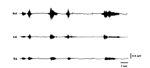 |
Fig. 1 Electromyogram patterns of lower leg muscles in a baby girl during the first bipedal walking without any supports in her life (at the age of 12 months and 11 days). Sol, LG, and TA:soleus, lateral portion of gastrocnemius, and tibialis anterior.
|
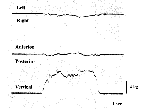 |
| Fig. 2 Three-dimensional ground reaction forces recorded in a baby girl during the first bipedal walking without any supports in her life. |
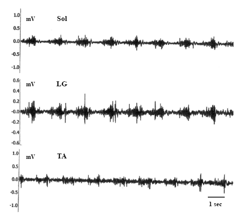 |
Fig. 3 Electromyogram patterns of lower leg muscles in a 15-year-old girl. Sol, LG, and TA:soleus, lateral portion of gastrocnemius, and tibialis anterior.
|
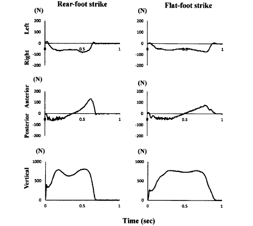 |
Fig. 4 Typical patterns of three-dimensional ground reaction forces recorded in an adult male subject during the regular rear-foot strike walking and mimicked flat-foot strike walking.
|
CONCLUSIONS
Co-contraction of ankle dorsi-flexors (soleus and LG) and plantar-flexor (TA) was observed during the first independent walking in a neonate. The EMG amplitudes and the step intervals were not constant. But disturbance of body balance toward both anterior/posterior and right/left directions was minor, although some degree of fluctuation was noted in the vertical force during the body-support phase. The relatively stable forces toward anterior and posterior direction suggested that her landing patterns were flat-foot strike without heel-strike and toe-kick. The co-contraction of antagonists disappeared and walking patterns were normalized at the age of 15. Since the afferent neural input is closely related to the antigravity activity of muscle, it was speculated that afferent input may play an important role in the learning of walking motor performance in new-born babies.
ACKNOWLEDGEMENTS
This study, in part, was supported by the Grant-in-Aid for Scientific Research (S-19100009, YO, http://www.jsps.go.jp/j-grantsinaid/index.html) from Japan Society for the Promotion of Science, Grant-in-Aid for Research Activity Start-up (TakO). The funders had no roles in the study design, data collection and analyses, decision to publish, or preparation of the manuscript. No additional external funding received for this study.
AUTHOR CONTRIBUTIONS
LO and TakO contributed to the analyses of data. KK and TomO performed the experiments using an infant and a girl, respectively. TW, KK, YN, and YO performed the experiment III. LO, TakO, and YO contributed to all of the aspects in this study, including drafting of the manuscript and executing the final revisions. All authors approved the final manuscript prior to publication.
FINANCIAL DISCLOSURE
Authors have no financial interest related to this project.
REFERENCES
| 1) |
Assaiante, C. and Chabrol, B.:Developmental and locomotor disorders in children. Revue Neurol. (Paris), 166, 149-157, 2010. |
| 2) |
Bosch, K. and Rosenbaum, D.:Gait symmetry improves in childhood - a 4-year follow-up of foot loading data. Gait & Posture, 32, 464-468, 2010.
|
| 3) |
Dominici, N., Ivanenko, Y.P., Cappellini, G., d’Avella, A., Mondi, V., Cicchese, M., Fabiano, A., Silei, T., Di Paolo, A., Giannini, C., Poppele, R.E. and Lacquaniti, F.:Locomotor primitives in newborn babies and their development. Science, 334, 997-999, 2011. |
| 4) |
Edgerton, V.R., McCall, G.E., Hodgson, J.A., Gotto, J., Goulet, C., Fleischmann, K. and Roy, R.R.:Sensorimotor adaptations to microgravity in humans. J. Exp. Biol., 204, 3217-3224, 2001.
|
| 5) |
Iosa, M., Fusco, A., Morone, G. and Paolucci, S.: Development and decline of upright gait stability. Front. Aging Neurosci., 6, 14, 2014. |
| 6) |
Ivanenko, Y.P., Dominici, N., Cappellini, G., Dan, B., Cheron, G. and Lacquaniti, F.:Development of pendulum mechanism and kinematic coordination from the first unsupported steps in toddlers. J. Exp. Biol., 207, 3797-3810, 2004.
|
| 7) |
Ivanenko, Y.P., Dominici, N., Cappellini, G., Di Paolo, A., Giannini, C., Poppele, R.E. and Lacquaniti, F.:Changes in the spinal segmental motor output for stepping during development from infant to adult. J. Neurosci., 33, 3025-3036, 2013.
|
| 8) |
Ivanenko, Y.P., Dominici, N., Cappellini, G. and Lacquaniti, F.: Kinematics in newly walking toddlers does not depend upon postural stability. J. Neurophysiol., 94, 754-763, 2005.
|
| 9) |
Kawano, F., Ishihara, A., Stevens, J.L., Wang, X.D., Ohshima, S., Horisaka, M., Maeda, Y., Nonaka, I. and Ohira, Y.: Tension- and afferent-input-associated responses of neuromuscular system of rats to hindlimb unloading and/or tenotomy. Am. J. Physiol. Regul. Integ. Com. Physiol., 287, R76-R86, 2004.
|
| 10) |
Kawano, F., Nomura, T., Ishihara, A., Nonaka, I. and Ohira, Y.:Afferent input-associated reduction of muscle activity in microgravity environment. Neuroscience, 114, 1133-1138, 2002.
|
| 11) |
Layne, C.S., Lange, G.W., Pruett, C.J., McDonald, P.V., Merkle, L.A., Mulavara, A.P., Smith, S.L., Kozlovskaya, I.B. and Bloomberg, J.J.:Adaptation of neuromuscular activation patterns during treadmill walking after long-duration space flight. Acta Astronaut., 43, 107-119, 1998.
|
| 12) |
Layne, C.S., Mulavara, A.P., Pruett, C.J., McDonald, P.V., Kozlovskaya, I.B. and Bloomberg, J.J.:The use of in-flight foot pressure as a countermeasure to neuromuscular degradation. Acta Astronaut., 42, 231-246, 1998.
|
| 13) |
Miller, C.A., Peters, B.T., Brady, R.R., Richards, J.R., Ploutz-Snyder, R.J., Mulavara, A.P. and Bloomberg, J.J.: Changes in toe clearance during treadmill walking after long-duration spaceflight. Aviat. Space Environ. Med., 81, 919-928, 2010.
|
| 14) |
Nielsen, J.B. and Sinkjaer, T.:Afferent feedback in the control of human gait. J. Electromyog. Kinesiol., 12, 213-217, 2002.
|
| 15) |
Ohira, T., Okabe, H., Kawano, F., Oke, Y., Fujita, R., Nomura, S., Nakai, N., Ohira, T., Ohira, K. and Ohira, Y.: Reconsideration of exercise prescription as the countermeasure for prevention of muscle atrophy in space. Experimental Biology 2010, Anaheim, California, U.S.A., April 24-28, No. 616.10, 2010.
|
| 16) |
Ohira, T., Terada, M., Kawano, F., Nakai, N., Ogura, A. and Ohira, Y.:Region-specific responses of adductor longus muscle to gravitational load-dependent activity in Wistar Hannover rats. PLoS ONE, 6, e21044, 2011.
|
| 17) |
Ohira, Y., Kawano, F., Wang, X.D., Sudoh, M., Iwashita, Y., Majima, H.J. and Nonaka, I.:Irreversible morphological changes in leg bone following chronic gravitational unloading of growing rats. Life Sci., 79, 686-694, 2006.
|
| 18) |
Ohira, Y., Nomura, T., Kawano, F., Sato, Y., Ishihara, A. and Nonaka, I.:Effects of nine weeks of unloading on neuromuscular activities in adult rats. J. Gravitat. Physiol., 9, 49-59, 2002.
|
| 19) |
Okamoto, T. and Okamoto, K.:Electromyographic characteristics at the onset of independent walking in infancy. Electromyog. Clin. Neurophysiol., 41, 33-41, 2001.
|
| 20) |
Okamoto, T., Okamoto, K. and Andrew, P.D.: Electromyographic developmental changes in one individual from newborn stepping to mature walking. Gait & Posture, 17, 18-27, 2003. |
| 21) |
Sala, D.A. and Cohen, E.:Gait component changes observed during independent ambulation in young children. Ind. J. Pediat., 80, 397-403, 2013. |
| 22) |
Sutherland, D.H., Olshen, R., Cooper, L. and Woo, S.L.: The development of mature gait. J. Bone Joint Surg. Am., 62, 336-353, 1980. |
| 23) |
Teulier, C., Sansom, J.K., Muraszko, K. and Ulrich, B.D.: Longitudinal changes in muscle activity during infants’ treadmill stepping. J. Neurophysiol., 108, 853-862, 2012. |
Send correspondence to:
1-3 Miyakodani, Tatara, Kyotanabe City, Kyoto 610-0394, Japan
Graduate School of Health and Sports Science, Doshisha University
Y. Ohira, Ph.D.
Phone:+81-774-65-7163
FAX:+81-774-65-6029
E-mail:yohira@mail.doshisha.ac.jp