宇宙航空環境医学 Vol. 48, No. 3, 23-34, 2011

原 著

Modulation of Hippocampal Proteins by Exposure to Simulated Microgravity Environment during the Postnatal Development in Rats

Yoshihiko Oke1, Fuminori Kawano1, Sachiko Nomura1, Takashi Ohira2, Ryo Fujita1, Masahiro Terada2, Tsubasa Shibaguchi2, Naoya Nakai1 and Yoshinobu Ohira1, 2

Graduate School of 1Medicine and 2Frontier Biosciences, Osaka University


ABSTRACT
 Effects of 14 days of hindlimb suspension and 14 days of ambulation recovery on the hippocampus proteome were investigated in 3-week old male Wistar Hannover rats during postnatal developing stage. Hindlimb suspension caused increased and decreased expression of 4 and 6 proteins, respectively. All of these proteins are reported to have functions related to cellular protection, cell death, neurogenesis, neural activity, or neuronal circuitry formation. In order to reveal whether post-translational modifications are altered, we further studied isoelectric point profiles of the 3 proteins (protein disulfide isomerase A3, triosephosphate isomerase 1, and sirtuin 2) involved in cell death or neuronal circuitry formation. Each of the 3 proteins indicated different spot patterns between the control and hindlimb-suspended groups. Furthermore, the altered profiles of proteins were not normalized to the age-matched control levels after 14 days of recovery. These results suggest that hindlimb suspension during the postnatal development of the brain might induce an inappropriate cell survival and abnormal neuronal circuitry, followed by disorder in the hippocampus function.

(Received : 12 May, 2011 Accepted : 8 February, 2012)

Key words : Microgravity, Hindlimb suspension, Proteome, Hippocampus, Postnatal developing rat

INTRODUCTION
 Exposure to actual and/or simulated microgravity environment causes various physiological adaptations, including in the central nervous system5,6,8,19) and antigravity muscle21,29) of human and rats. For example, 16 days of spaceflight leads to a change in the synaptic circuitry at the hindlimb cortex of the postnatal developing rats6). Therefore, understanding of the mechanism responsible for the molecular adaptation to inhibited antigravity activity in space or during prolonged bed rest is important.
 The hindlimb suspension model in rats is widely used to study the effects of simulated microgravity environment. This model induces a redistribution of fluid in the body7) and atrophy of soleus muscle, an antigravity hindlimb muscle23,38). In terms of the nervous system, the hindlimb suspension causes the decrease of electromyogram activity in soleus muscle, afferent neurogram in spinal cord22), GABAergic neurons in the hindlimb somatosensory cortex5), and neurogenesis in rats41). Recently, proteomic analyses of the hippocampus34) and the hypothalamus35), as well as a microarray gene expression analysis of mouse brain11), were performed to elucidate the mechanism responsible for the adaptation to gravitational unloading. After 7 days of hindlimb suspension, the expression of cytoskeletal proteins, such as tubulin, and metabolic proteins in hippocampus of adult mice was changed (7 spots were decreased and 4 spots were increased)34), and biomarkers of oxidative stress in hypothalamus of mice were increased35). Therefore, exposure to simulated microgravity environments by the hindlimb suspension might induce distinct changes specific to the regions of the brain or the different species.
 The hippocampus is an important brain region to integrate the information arriving from different sensory organs and to relate with memory formation. On the postsynaptic density in the hippocampus, protein composition, such as the developmental switch in N-methyl-D-aspartate (NMDA) receptor type and the receptor-associated proteins, changes during the early development in rats from postnatal day 2 to 3532). These alternations contribute to the maturation of learning and memory. Hence, there is a possibility that the development of rat hippocampus could be susceptible to microgravity during the postnatal developing stage of the brain. However, it remains unclear how antigravity activity of muscles influence the postnatal development of brain. Therefore, this study was performed to investigate the effects of hindlimb suspension during postnatal development on both expression levels and post-translational modifications of hippocampal proteins in rats. Effects of ambulation recovery were also investigated.

MATERIALS AND METHODS
 A. Animal care and experimental procedures
 All experimental procedures were conducted in accordance with the Japanese Physiological Society Guide for the Care and Use of Laboratory Animals and followed the guiding principles of the American Physiological Society. This study was also approved by the Committee on Animal Care and Use at Osaka University.
 Male Wistar Hannover rats (Nihon CLEA, Tokyo, Japan) with the age of 3-week old were used in this study. The rats were randomly separated into cage-control (n=15) and hindlimb-suspended group (n=10). Each rat was housed in a cage (30×30 cm with 30 cm height). Temperature and humidity in the animal room with 12 : 12 hr light : dark cycle were maintained at 〜23°C and 〜55%, respectively. Solid food (CE-2, Nihon CLEA) and water were supplied ad libitum.
 On the first day of the experiment, brains were sampled from 5 rats anesthetized with intraperitoneal injection of pentobarbital sodium (5 mg/100 g body weight) after perfusion using phosphate buffered saline to remove the blood from the brain and these samples served as the pre-experimental controls. Hippocampus was sampled, minced, frozen in liquid nitrogen, and stored at −80°C. The hindlimb suspension was performed as described previously28). Breifly, three pieces of sticky tape with good cushion were placed longitudinally on 3 sides of the mid-tail of the rat. The attached tapes were loosely wrapped with another tape cross-sectionally to keep the blood flow intact. A string was inserted through the gap between the tail and sticky tapes. The string was fastened to a horizontal bar on the top of a plastic cage (30×30 cm and 30 cm height) and manipulated to elevate the hindlimbs to avoid touching the floor and walls of the cage. The rats could reach food and water freely using their forelimbs. After 14 days, both cage control and the hindlimb-suspended rats (n=5 in each group) were sacrificed and the hippocampus was sampled. Ambulation recovery was allowed for the remaining rats and hippocampus was sampled after 14 days.
 B. Preparation of hippocampal protein extracts
 Frozen hippocampus was powdered in stainless steel mortar cooled by liquid nitrogen and suspended in lyses buffer (20 mM Tris-HCl with pH 7.6, 150 mM NaCl, 50 mM NaF, 1 mM Na3VO4, 1% Triton X-100, and 1% protease inhibitor cocktail I (Millipore, MA, USA). The suspension was gently rotated to extract proteins from powdered tissue for 16 h at 4°C and centrifuged at 20,000 ×g for 10 min. The supernatant was filtered with Ultrafree-MC centrifugal filter units (Millipore). The protein content in the extracts was determined using BCA Protein Assay Kit (Thermo Scientific, MA, USA).
 C. 2-Dimensional gel electrophoresis (2-DE)
 Equal amount (110 µg) of hippocampal protein extracted from 3 rats at day 14 was mixed (total 330 µg for each group). Each mixture was desalted by using 2-D Clean-Up Kit (GE Healthcare, Buckinghamshire, UK) and dissolved in DeStreak Rehydration Solution with 2% IPG buffer for pH 3-10 NL (GE Healthcare). The samples were applied on immobilized pH 3-10 nonlinear gradient dry strip gels (7 cm, GE Healthcare). After 12 h rehydration, the isometric focusing was performed at constant 500 V for 1 h (20°C) in Ettan IPGphor II (GE Healthcare). The voltage was then raised to 1,000 V for 1 h and finally to 5,000 V for 3 h. The focused strips were washed twice in deionized distilled water for 1 h (total 2 h) and equilibrated for 15 min in 50 mM Tris-HCl with pH 8.8, 6 M urea, 30% glycerol, 2% sodium dodecyl sulfate (SDS), and 0.05% bromophenol blue, containing 10 mg/mL dithiothreitol, followed by 25 mg/mL iodoacetamide in the same buffer for 15 min at 23°C. The second-dimensional separation was performed on 12.5% SDS-polyacrylamide gel electrophoresis (SDS-PAGE). The gels were stained with coomassie brilliant blue R-250 for 30 min and the excess dye was washed out from the gels by destaining with 20% methanol and 5% acetic acid. Molecular masses were determined by running standard protein marker (Bio-Rad laboratories, CA, USA) and isoelectric point (pI) values were provided by the supplier of the dry strip gels. The experiments were repeated 3 times and the results were reproducible.
 D. In-gel digestion and peptide fragments
 Spots were excised, destained with 50% methanol in 50 mM ammonium bicarbonate at 40°C, and dried by reduced-pressured centrifugation. Each piece of dried gel was rehydrated with 20 µL of 50 mM Tris-HCl (pH 8.8) containing 1 pmol trypsin (Promega, WI, USA) for 10 min at 4°C. After removal of excess rehydrate solution, 15 µL of 50 mM Tris-HCl (pH 8.8) were added to the gel piece and the mixture was incubated for 10 h at 37°C. Then, 100 µL of 0.1% trifluoroacetic acid (TFA) were added to the mixture. The peptide fragments were collected as the supernatant and further eluted from the residue of the gel by 10 min-sonication (twice) in 100 µL of 50% acetonitrile (ACN) containing 0.1% TFA, followed by 10-min sonication (once) in 80% ACN with 0.1% TFA. The mixture of the eluted peptides was filtered with Ultrafree-MC centrifugal filter units and concentrated by reduced-pressured centrifugation. Next, the peptides were desalted using C18 ZipTips (Millipore). The ZipTip was pre-wet by aspirating 10 µL of 80% ACN with 0.1% TFA (3 times) and 10 µL of 0.1% TFA (twice). Then, the peptides were absorbed to ZipTip by pipetting 20 times. After the ZipTip was washed with 0.1% TFA, the peptides were eluted in 50% ACN with 0.1% TFA. The eluate was diluted with 10 µL of 0.1% TFA, and ACN was evaporated by reduced-pressured centrifugation.
 E. Matrix-assisted laser desorption ionization time-of-flight mass spectrometry (MALDI-TOF/MS)
 Each purified peptide fraction (1 µL) was mixed with the matrix solution (1 µL) containing α-cyano-4-hydroxycinnamic acid (Bruker Daltonics) in 33% ACN with 0.06% TFA, and the mixture (2 µL) was applied onto a target plate (MTP 384 target plate ground steel TF, Bruker Daltonics, MA, USA). The samples were analyzed with peptide mass fingerprinting by using Ultraflex mass spectrometry (Bruker Daltonics). MASCOT search in the NCBI database was performed with the obtained mass of peptides.
 F. Western blotting
 Western blotting was performed as described previously23). Briefly, hippocampal protein extracts were dissolved in equal volumes of 2×SDS sample buffer (20% glycerol, 12% 2-mercaptoethanol, 4% SDS, 100 mM Tris-HCl with pH 6.7, 0.05% bromophenol blue) and adjusted to a final concentration of 0.4 (for triosephosphate isomerase 1, TPI1), 2 (for protein disulfide-isomerase associated 3, PDIA3), or 0.16 µg/µL (for sirtuin 2, SIRT2) by 1 SDS sample buffer.
 All samples of 10 µL were separated by 10% SDS-PAGE and transferred to polyvinylidene difluoride (PVDF) membranes (Bio-Rad laboratories). The membranes were blocked in 5% skimmed milk (Nacalai Tesque, Kyoto, Japan) with 0.1% Tween 20 in Tris-buffered saline (TTBS) for 1 h at 23°C and incubated with anti-TPI1 (1:30,000, Abnova, Taipei City, Taiwan), anti-PDIA3 (1:2,000, Abnova) or anti-SIRT2 (1:2,000, Cell Signaling Technology, MA,USA) in TTBS containing 5% bovine albumin (Nacalai Tesque) for 12 h at 4°C. Then, the blots were incubated with horseradish peroxidase-conjugated secondary antibody to goat (1:5,000, Santa Cruz Biotechnology, CA, USA) or rabbit immunoglobulin G (1:5,000, Cell Signaling Technology) for 1 h. The proteins reacted with antibodies were detected by incubation of the blots with ECL plus Kit (GE Healthcare) and followed by exposing to panchromatic film (FP-3000B, Fuji Photo Film, Tokyo, Japan). After the analyses, the bound antibodies were stripped from the membranes by 15 min-incubation in stripping solution (Nacalai Tesque) at 23°C. The membranes were blocked again and incubated with anti-β-actin (1:5,000, Santa Cruz Biotechnology) in TTBS containing 5% skimmed milk for 12 h at 4°C. The blots were incubated with horseradish peroxidase-conjugated secondary antibody to mouse immunoglobulin G (1:5,000, Cell Signaling Technology) for 1 h. The detection of the bands was performed as was stated above. The analyses were performed in 5 rats within each group, and the results were reproducible.
 For 2-DE western blotting analysis of TPI1, PDIA3 and SIRT2, hippocampal protein extracts from 5 rats in each group were mixed and adjusted to 2, 5, and 16 µg protein, respectively. Each mixture was desalted and dissolved in DeStreak Rehydration Solution with 0.5% IPG buffer for pH 3-10 NL. All of the 2-DE western blotting analyses were repeated twice, and the results were reproducible.

RESULTS
 A. 2-DE analysis of hippocampal proteins
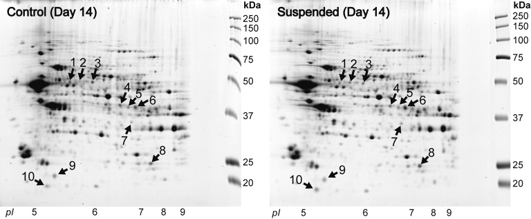
 To investigate whether the hindlimb suspension during the postnatal development affects the protein expression profile in the hippocampus of rat, 2-DE maps of overall proteins were compared between the control and hindlimb-suspended group. Representative 2-DE maps of 2 groups are shown in Figure 1. Differentially-displayed proteins in the hippocampus of rats after 14 days of hindlimb suspension were examined by visual map matching. In comparison with the control group, 4 weaker spots and 6 stronger spots were detected in the hindlimb-suspended group (Fig. 1 and Table 1). All of the 10 spots were identified by peptide mass fingerprinting and the results including their Molecular Weight Search scores are shown in Table 1. The levels of dihydropyrimidinase-like 2 (DPYSL2), actin-related protein 1 homolog A, centractin alpha, and phosphatidylethanolamine binding protein (PEBP) in the hindlimb-suspended group were lower than controls. In contrast, the levels of SIRT2, TPI1 and peroxiredoxin 2 (PRDX2) in the hindlimb-suspended group were higher than controls. PDIA3 (Spot 2 and 3 in Fig. 1 and Table 1) and glutamine synthetase 1 (Glul) (Spot 5 and 6 in Fig. 1 and Table 1) were respectively detected as two spots. The PDIA3 spot with a lower pI (Spot 2 in Fig. 1 and Table 1) increased and the spot with a higher pI decreased by hindlimb suspension, on the contrary (Spot 3 in Fig. 1 and Table 1). The densities of both Glul spots were higher in the hindlimb-suspended group than controls (Spot 5 and 6 in Fig. 1 and Table 1).

Fig. 1. Representative 2-dimensional gel electrophoresis maps of hippocampal proteins in control and suspended rats just after the 14 days of cage housing or hindlimb suspension. Differentially expressed protein spots between the control and suspended rats are marked with arrows and numbers. pI, isoelectric point.


Table 1. Alternation in hippocampal protein expressions by the tail-suspension
Spot # Protein name Accession # Predicted MW (kDa) MOWSE Score Δ (Spot of Sus vs Cont at R+0)
1 Dihydropyrimidinase-like 2 (DPYSL2) gi|40254595 63 83 Down
2 Protein disulfide-isomerase A3 (PDIA3) gi|1352384 57 127 Up
3 Protein disulfide-isomerase A3 (PDIA3) gi|8393322 57 201 Down
4 Actin-related protein 1 homolog A, centractin alpha gi|5031569 42 86 Down
5 Glutamine synthetase 1 (Glul) gi|142349612 43 168 Up
6 Glutamine synthetase 1 (Glul) gi|142349612 43 212 Up
7 Sirtuin 2 (SIRT2) gi|56605812 40 128 Up
8 Triosephosphate isomerase 1 (TPI1) gi|117935064 27 328 Up
9 Phosphatidylethanolamine binding protein (PEBP) gi|8393910 21 108 Down
10 Peroxiredoxin 2 (PRDX2) gi|8394432 22 95 Up
All spot numbers are consistent with the numbers described in Figure 1.

 B. Western blotting analyses of protein disulfide-isomerase associated 3, triosephosphate isomerase 1 and Sirtuin 2
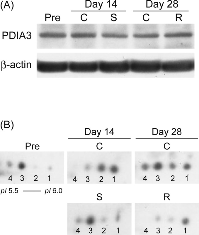
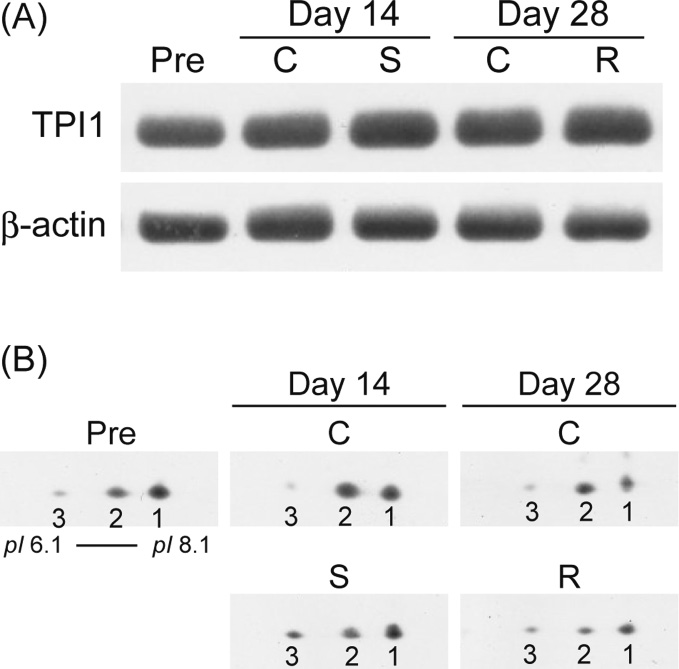
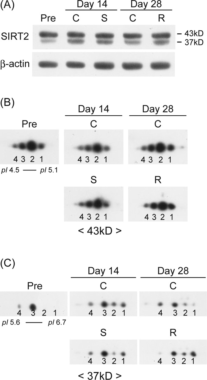
 There were not significant differences in the total expression levels of PDIA3, TPI1, and SIRT2 among all groups, except the 37 kD-form SIRT2, which intensities were significantly increased at day 14 and 28 vs. pre-experimental basal level, respectively (Figs. 2A, 3A, and 4A). However, by using 2-DE western blotting analysis, the data suggested that the pI profiles of TPI1, PDIA3, and 37 kD-form SIRT2 could be greatly influenced by the hindlimb suspension (Figs. 2B, 3B, and 4C). As is shown in Figure 2B, 4 spots with a different pI were detected as PDIA3 by using 2-DE western blotting. Before the initiation of experiment, 2 spots of PDIA3 with a lower pI were clearly observed (Spot 3 and 4), but other 2 spots with a higher pI were hardly detected (Spot 1 and 2). In the control group at day 14, 2 spots with a higher pI were clearly noted and other 2 spots with a lower pI almost disappeared. In the control group at day 28, the 2 spots with a lower pI were clearly observed again. Interestingly, the patterns of PDIA3 spots in the hindlimb-suspended group at day 14 were similar to those seen before experiment. Those in the ambulation recovery group at day 28 were similar to those in the control group at day 14.
 As for TPI1, 3 spots with different pI were detected (Fig. 3B). At the age of 3 week, the greatest expression was noted in the spot with the highest pI (Spot 1). In the control groups both at day 14 and 28, the greatest expression was observed in the spot with the second highest pI (Spot 2). However, in the hindlimb-suspended group at day 14 and 28, the order of expression levels was similar to the pre-experimental controls.
 SIRT2 showed 2 isoforms, 43 kD-form and 37 kD-form (Fig. 4A). The pI profile of 43 kD-form SIRT2 was not influenced by the hindlimb suspension or the postnatal growth during week 3 to 7 after birth (Fig. 4B). On the other hand, the pI profile of 37 kD-form SIRT2 changed in response to the postnatal development and hindlimb suspension (Fig. 4C). In 3-week old rats, only 2 spots of 37 kD-form SIRT2 with lower pI were detected (Spot 3 and 4). In the control group at day 14, additional 2 spots with higher pI appeared, but these 2 spots, especially with the highest pI, decreased during the next 14 days (Spot 1 and 2). During the postnatal development of the brain, the expression of the spot with the lowest pI (No. 4) increased gradually in the control group. However, 2 spots with higher pI increased progressively in response to 14 days of unloading and reloading in the experimental group. At the end of ambulation recovery, the spot with the lowest pI became negligible.

Fig. 2. Expression level and 2-dimensional gel electrophoresis (2-DE) profile of protein disulfide-isomerase associated 3 (PDIA3) in the hippocampus. (A) The representative 1-DE western blotting patterns of PDIA3 are shown. (B) PDIA3 protein spots were verified by 2-DE western blotting analysis. Spots of PDIA3 are numbered beginning at the highest isoelectric point. Pre, pre-suspension control ; Day 14, 14 days after cage housing (C) or hindlimb suspension (S) ; Day 28, 28 days after cage housing (C) or 14 days after ambulation recovery following 14-day hindlimb suspension (R). See figure 1 for other abbreviation.
 
Fig. 3. Expression level and 2-DE profile of triosephosphate isomerase 1 (TPI1) in the hippocampus. (A) The representative 1-DE western blotting analysis of TPI1 is shown. (B) TPI1 protein spots were verified by 2-DE western blotting analysis. Spots of TPI1 are numbered beginning at the highest isoelectric point. See figure 1 and 2 for other abbreviations.
 
Fig. 4. Expression level and 2-DE profile of Sirtuin 2 (SIRT2) in the hippocampus. (A) The representative 1-DE western blotting patterns of SIRT2 are shown. SIRT2 protein spots with 43 kD (B) and 37 kD (C) were respectively verified by 2-DE western blotting analysis. Spots of SIRT2 with either 43 kD or 37 kD are numbered beginning at the highest isoelectric point. See figure 1 and 2 for other abbreviations.


DISCUSSION
 Gravitational unloading during the postnatal development inhibited the normal growth-associated changes of hippocampal protein expression in rats. Further, 14 days of ambulation recovery from the hindlimb suspension could not restore the hippocampal protein profiles to the age-matched control level. To our knowledge, this is the first report concerning the effects of gravitational unloading and reloading on the hippocampal expression of proteins in the postnatal developing rats, although the effects of hindlimb suspension on the hippocampal protein profiles in adult mice were reported elsewhere34).
 The hippocampal proteins, which showed different expressions between the control and the hindlimb-suspended group at day 14, have functions related to cellular protection, cell death, neurogenesis, neural activity or neuronal circuitry formation, as will be described in detail below. The data suggested that inhibition of antigravity activity of hindlimbs during early growing period might impair the normal postnatal development and normal function of the hippocampus.
 A. Cell survival ─ cellular protection, cell death and neurogenesis ─
 The hindlimb suspension of mice causes oxidative stress in the brain and lipid peroxidation in the hippocampus39). Hence, various reactions, such as adaptive response to the stress, cell dysfunction or cell death, may occur in the hippocampus of the hindlimb-suspended mice and rats. Furthermore, the hindlimb suspension suppresses neurogenesis in the hippocampus of the postnatal developing rats27), as well as adult rats41).
 The up-regulation of PRDX2 and the change of post-translational modifications of PDIA3 might be induced in response to cellular stresses in the hippocampus. PRDX2 contributes to elimination of peroxides. Actually, over-expression of PRDX2 rescues PC12 cells and cortical neuronal culture from cell death caused by oxidative injury2,37). PRDX2 may also play an important role in the defense against neurodegenerative diseases, because the expression levels of PRDX2 increase in postmortem brain of patients with Down’s syndrome, Alzheimer’s, Parkinson’s, and Pick’s diseases, in which oxidative stress is a common element24). Therefore, it is speculated that PRDX2 might be up-regulated to defend hippocampal cells against oxidative stress, when the hindlimb suspension was performed in the postnatally developing rats. Responses of other stress markers, other than PRDX2, were not investigated in the present study.
 PDIA3 assists the chaperon function and disulfide shuffling of glycoproteins16). In addition, PDIA3 may act as a neuroprotective factor against neurotoxicity at an early stage of cell damage17). In Neuro-2a (N2a) neuroblastoma cells, siRNA inhibition of PDIA3 significantly enhances prion toxicity, which induces neuronal apoptosis via activation of the endoplasmic reticulum stress pathway, and over-expression of PDIA3 protects cells against the toxicity by reduction of caspase-12 activity17). Moreover, the pI of PDIA3 in lung tissue shifts to lower level after exposure of neonatal rats to hyperoxic condition, in which apoptotic cell death of lung cells can be induced by reactive oxygen species40). Similar PDIA3 spot pattern between the hindlimb-suspended group at day 14 (Fig. 2B) and the hyperoxia-exposed lung tissue was observed. Thus, the hindlimb suspension might trigger apoptotic cell death via cell stress in the hippocampus. Furthermore, similarities of spot patterns were noted between the ambulation recovery group at day 28 and the control group at day 14, as well as the hindlimb-suspended group at day 14 and the pre-experimental controls. It is suggested that the hindlimb suspension could temporarily inhibit the growth-related change of post-translational modifications of PDIA3, but this inhibition could be reversible after the termination of hindlimb suspension.
 There are relationships between cell death and cellular functions in several degenerative diseases14,36). The expression of centractin is down-regulated in Down’s syndrome patients14) and mutations of TPI1 cause triosephosphate isomerase enzymopathy, which is a unique glycolytic enzyme deficiency coupled with neurodegeneration36). Centractin mediates the retrograde axonal transport of neurotrophines33) and acts as a regulator for various dynein-mediate functions during mitosis10). Therefore, reduction of centractin in the hindlimb-suspended rats at day 14 might be implicated in an increase of cell death or a decrease of the neurogenesis (Fig. 1, Spot 4). However, prominent unloading-related changes were not noted in the volume of hippocampus.
 TPI1 is inactivated by cyclinA/Cdk2-dependent phosphorylation25) or nitrotyrosination from amyloid β-peptide-induced nitro-oxidative damage13). The phosphorylation of TPI1 occurs during etoposide-induced apoptosis and the nitro-TPI1 induces tau aggregation to form paired helical filaments, the characteristic intracellular hallmark of brain in patient with Alzheimer’s disease. Generally, these post-translational modifications shift the pI of protein to lower level. Then, shifts of pI of TPI1 toward the lower level are considered to lead to cell death. Since TPI1 spots with lower pI increased in all groups at day 14 and 28 as compared to the pre-experiment control (Fig. 3, Spot 2 in the control group and Spot 3 in the hindlimb-suspended group and the ambulation recovery group), the rates of cell death might be increased in these groups at day 14 and 28, although the molecular manners for the death might be different between the control and the hindlimb-suspended groups. In addition, since the spot patterns of TPI 1, which were altered by hindlimb suspension, was maintained even after the 2-week ambulation recovery, it is suggested that the inhibited post-translational modifications may not be restored to the age-matched control level.
 B. Neurotransmission ─ neuronal circuitry and neural activity ─
 DPYSL2 is a key molecule for axon guidance and neuronal growth cone collapse18,42). Dissociation of DPYSL2 from tubulin after its phosphorylation leads to induction of growth cone collapse4,42). On the other hand, hyper-phosphorylation of DPYSL2 is implicated in some pathology, such as Alzheimer’s disease4). Furthermore, DPYSL2 expression decreases in the frontal cortex of psychiatric patients, such as schizophrene20). Therefore, less expression of DPYSL2 after hindlimb suspension might indicate that formation of normal neuronal circuitry in the hippocampus is inhibited and subsequently cognitive function may be impaired.
 The expression of PEBP mRNA in the brain of Alzheimer’s disease patients decreases, particularly in the hippocampal CA1 field of patients with late-onset Alzheimer’s disease26). And the decrease in PEBP protein expression correlates with amyloid β accumulation in the mouse model of Alzheimer’s disease12). PEBP is the precursor of the hippocampal cholinergic neurostimulating peptide, which increases the production of choline acetyltransferase30). Recently, reduction in rat PEBP, which is paralleled with cognitive impairment after chronic corticosterone treatment, is reported9). Therefore, reduction of PEBP in the hippocampus after hindlimb suspension may cause cholinergic impairment and cognitive dysfunction.
 Glul is exclusively expressed in astrocyte and plays a central role in the metabolic regulation of glutamate, the major excitatory brain neurotransmitter1). Glutamate is taken up by astrocytes through the astrocyte-specific glutamate transporter-1 and transformed into glutamine by Glul. It is reported that the increase in hippocampal Glul activity and the mild impairment of memory formation are induced by chronic exposure to glufosinate-ammmonium, which is an inhibitor of glutamine synthetase in plants3). Thus, higher expression of Glul in hindlimb-suspended animal at day 14 suggests that the hindlimb suspension might induce a disturbance of the glutamate-glutamine cycle in the hippocampus and an impairment of memory formation.
 SIRT2 is a multifunctional brain protein, which can inhibit cellular events implicated in neuronal circuitry formation, such as cytoskeletal growth cone dynamics, neurite outgrowth, and oligodendrocyte arborization15,31). The activated SIRT2 also enhances neurodegeneration15). SIRT2 is inactivated by its phosphorylation and acetylation, which are induced by various cyclin-Cdk complexs and p300/CBP. Hindlimb suspension affected only pI profile of 37 kD-form SIRT2 in the present study. The expression of 37 kD-form SIRT2 with highest pI (Fig. 4C, Spot 1) was stimulated by suspension. That in 3-week old control rats was not detected, although it was increased at day 14. However, in the hindlimb-suspended group with or without ambulation recovery, the expression was increased. Therefore, SIRT2-dependent neurodegeneration or inhibition of cellular events related with neuronal circuitry formation might be enhanced at both postnatal week 5 with hindlimb suspension and week 7 after the ambulation recovery, although those might be enhanced only around postnatal week 5 under normal condition. Hence, the hindlimb suspension during postnatal development of the brain may have a risk of neurodegeneration or disturbance of neuronal circuitry formation.

CONCLUSION
 Taken together, it was suggested that hindlimb unloading might trigger cell death signal via cell stress or form an abnormal neuronal circuitry in the hippocampus of the postnatal developing brain, which might cause adverse effects on hippocampal function. In addition, there is a possibility that the effects caused by hindlimb suspension might be maintained for long periods or irreversible, because the spot patterns of TPI1 and SIRT2 were not normalized to the age-matched control levels after 14 days of ambulation. Although it remains unknown whether these alternations of protein profiles would be beneficial or not for the brain function, the results obtained in this study provided some new insights that post-translational modifications, as well as expression levels of proteins, are changed in the hippocampus of the postnatal developing rats in response to inhibited gravitational unloading.

ACKNOWLEDGEMENTS
 This study was supported by Grant-in-Aid for Scientific Research S (19100009) from Japan Society for the Promotion of Sciences and Grant-in-Aid for Young Scientists B (21700655) from the Ministry of Education, Culture, Sports, Science and Technology.


References

1) Bak, L.K., Schousboe, A. and Waagepetersen, H.S. : The glutamate/GABA-glutamine cycle : aspects of transport, neurotransmitter homeostasis and ammonia transfer. J. Neurochem., 98, 641-653, 2006.
2) Boulos, S., Meloni, B.P., Arthur, P.G., Bojarski, C. and Knuckey, N.W. : Peroxiredoxin 2 overexpression protects cortical neuronal cultures from ischemic and oxidative injury but not glutamate excitotoxicity, whereas Cu/Zn superoxide dismutase 1 overexpression protects only against oxidative injury. J. Neurosci. Res., 85, 3089-3097, 2007.
3) Calas, A.G., Richard, O., Meme, S., Beloeil, J.C., Doan, B.T., Gefflaut, T., Meme, W., Crusio, W.E., Pichon, J. and Montecot, C. : Chronic exposure to glufosinate-ammonium induces spatial memory impairments, hippocampal MRI modifications and glutamine synthetase activation in mice. Neurotoxicology, 29, 740-747, 2008.
4) Cole, A.R., Knebel, A., Morrice, N.A., Robertson, L.A., Irving, A.J., Connolly, C.N. and Sutherland, C. : GSK-3 phosphorylation of the Alzheimer epitope within collapsin response mediator proteins regulates axon elongation in primary neurons. J. Biol. Chem., 279, 50176-50180, 2004.
5) D’Amelio, F., Fox, R.A., Wu, L.C. and Daunton, N.G. : Quantitative changes of GABA-immunoreactive cells in the hindlimb representation of the rat somatosensory cortex after 14-day hindlimb unloading by tail suspension. J. Neurosci. Res., 44, 532-539, 1996.
6) DeFelipe, J., Arellano, J.I., Merchan-Perez, A., Gonzalez-Albo, M.C., Walton, K. and Llinas, R. : Spaceflight induces changes in the synaptic circuitry of the postnatal developing neocortex. Cereb. Cortex, 12, 883-891, 2002.
7) De Santo, N.G., Christensen, N.J., Drummer, C., Kramer, H.J., Regnard, J., Heer, M., Cirillo, M. and Norsk, P. : Fluid balance and kidney function in space : introduction. Am. J. Kidney Dis., 38, 664-667, 2001.
8) Edgerton, V.R., McCall, G.E., Hodgson, J.A., Gotto, J., Goulet, C., Fleischmann, K. and Roy, R.R. : Sensorimotor adaptations to microgravity in humans. J. Exp. Biol., 204, 3217-3224, 2001.
9) Feldmann, R.E. Jr., Maurer, M.H., Hunzinger, C., Lewicka, S., Buergers, H.F., Kalenka, A., Hinkelbein, J., Broemme, J.O., Seidler, G.H., Martin, E. and Plaschke, K. : Reduction in rat phosphatidylethanolamine binding protein-1 (PEBP1) after chronic corticosterone treatment may be paralleled by cognitive impairment : a first study. Stress, 11, 134-147, 2008.
10) Ford, J.H. : Spindle microtubular dysfunction in mothers of Down syndrome children. Hum. Genet., 68, 295-298, 1984.
11) Frigeri, A., Iacobas, D.A., Iacobas, S., Nicchia, G.P., Desaphy, J.F., Camerino, D.C., Svelto, M. and Spray, D.C. : Effect of microgravity on gene expression in mouse brain. Exp. Brain Res., 191, 289-300, 2008.
12) George, A.J., Holsinger, R.M., McLean, C.A., Tan, S.S., Scott, H.S., Cardamone, T., Cappai, R., Masters, C.L. and Li, Q.X. : Decreased phosphatidylethanolamine binding protein expression correlates with Abeta accumulation in the Tg2576 mouse model of Alzheimer’s disease. Neurobiol. Aging, 27, 614-623, 2006.
13) Guix, F.X., Ill-Raga, G., Bravo, R., Nakaya, T., de Fabritiis, G., Coma, M., Miscione, G.P., Villa-Freixa, J., Suzuki, T., Fernandez-Busquets, X., Valverde, M.A., de Strooper, B. and Munoz, F.J. : Amyloid-dependent triosephosphate isomerase nitrotyrosination induces glycation and tau fibrillation. Brain, 132, 1335-1345, 2009.
14) Gulesserian, T., Kim, S.H., Fountoulakis, M. and Lubec, G. : Aberrant expression of centractin and capping proteins, integral constituents of the dynactin complex, in fetal down syndrome brain. Biochem. Biophys. Res. Commun., 291, 62-67, 2002.
15) Harting, K. and Knoll, B. : SIRT2-mediated protein deacetylation : An emerging key regulator in brain physiology and pathology. Eur. J. Cell. Biol., 89, 262-269, 2010.
16) Hebert, D.N. and Molinari, M. : In and out of the ER : protein folding, quality control, degradation, and related human diseases. Physiol. Rev., 87, 1377-1408, 2007.
17) Hetz, C., Russelakis-Carneiro, M., Walchli, S., Carboni, S., Vial-Knecht, E., Maundrell, K., Castilla, J. and Soto, C. : The disulfide isomerase Grp58 is a protective factor against prion neurotoxicity. J. Neurosci., 25, 2793-2802, 2005.
18) Inagaki, N., Chihara, K., Arimura, N., Menager, C., Kawano, Y., Matsuo, N., Nishimura, T., Amano, M. and Kaibuchi, K. : CRMP-2 induces axons in cultured hippocampal neurons. Nat. Neurosci., 4, 781-782, 2001.
19) Ishihara, A., Ohira, Y., Roy, R.R., Nagaoka, S., Sekiguchi, C., Hinds, W.E. and Edgerton, V.R. : Effects of 14 days of spaceflight and nine days of recovery on cell body size and succinate dehydrogenase activity of rat dorsal root ganglion neurons. Neuroscience, 81, 275-279, 1997.
20) Johnston-Wilson, N.L., Sims, C.D., Hofmann, J.P., Anderson, L., Shore, A.D., Torrey, E.F. and Yolken, RH. : Disease-specific alterations in frontal cortex brain proteins in schizophrenia, bipolar disorder, and major depressive disorder. The Stanley Neuropathology Consortium. Mol. Psychiatry, 5, 142-149, 2000.
21) Kawano, F., Goto, K., Wang, X.D., Terada, M., Ohira, T., Nakai, N., Yoshioka, T. and Ohira, Y. : Role(s) of gravitational loading during developing period on the growth of rat soleus muscle fibers. J. Appl. Physiol., 108, 676-685, 2010.
22) Kawano, F., Ishihara, A., Stevens, J.L., Wang, X.D., Ohshima, S., Horisaka, M., Maeda, Y., Nonaka, I. and Ohira, Y. : Tension- and afferent input-associated responses of neuromuscular system of rats to hindlimb unloading and/or tenotomy. Am. J. Physiol. Regul. Integr. Comp. Physiol., 287, R76-86, 2004.
23) Kawano, F., Matsuoka, Y., Oke, Y., Higo, Y., Terada, M., Wang, X.D., Nakai, N., Fukuda, H., Imajoh-Ohmi, S. and Ohira, Y. : Role(s) of nucleoli and phosphorylation of ribosomal protein S6 and/or HSP27 in the regulation of muscle mass. Am. J. Physiol. Cell Physiol., 293, C35-44, 2007.
24) Krapfenbauer, K., Engidawork, E., Cairns, N., Fountoulakis, M. and Lubec, G. : Aberrant expression of peroxiredoxin subtypes in neurodegenerative disorders. Brain Res., 967, 152-160, 2003.
25) Lee, W.H., Choi, J.S., Byun, M.R., Koo, K.T., Shin, S., Lee, S.K. and Surh, Y.J. : Functional inactivation of triosephosphate isomerase through phosphorylation during etoposide-induced apoptosis in HeLa cells : Potential role of Cdk2. Toxicology, 278(2), 224-228, 2010.
26) Maki, M., Matsukawa, N., Yuasa, H., Otsuka, Y., Yamamoto, T., Akatsu, H., Okamoto, T., Ueda, R. and Ojika, K. : Decreased expression of hippocampal cholinergic neurostimulating peptide precursor protein mRNA in the hippocampus in Alzheimer disease. J. Neuropathol. Exp. Neurol., 61, 176-185, 2002.
27) Nomura, S., Kami, K., Kawano, F., Oke, Y., Nakai, N., Ohira, T., Fujita, R., Terada, M., Imaizumi, K. and Ohira, Y. : Effects of hindlimb unloading on neurogenesis in the hippocampus of newly weaned rats. Neurosci. Lett., in press.
28) Ohira, Y., Nomura, T., Kawano, F., Sato, Y., Ishihara, A. and Nonaka, I. : Effects of nine weeks of unloading on neuromuscular activities in adult rats. J. Gravit. Physiol., 9, 49-59, 2002.
29) Ohira, Y., Yoshinaga, T., Ohara, M., Nonaka, I., Yoshioka, T., Yamashita-Goto, K., Shenkman, B.S., Kozlovskaya, I.B., Roy, R.R. and Edgerton, V.R. : Myonuclear domain and myosin phenotype in human soleus after bed rest with or without loading. J. Appl. Physiol., 87, 1776-1785, 1999.
30) Ojika, K., Mitake, S., Kamiya, T., Kosuge, N. and Taiji, M. : Two different molecules, NGF and free-HCNP, stimulate cholinergic activity in septal nuclei in vitro in a different manner. Brain Res. Dev. Brain Res., 79, 1-9, 1994.
31) Pandithage, R., Lilischkis, R., Harting, K., Wolf, A., Jedamzik, B., Luscher-Firzlaff, J., Vervoorts, J., Lasonder, E., Kremmer, E., Knoll, B. and Luscher, B. : The regulation of SIRT2 function by cyclin-dependent kinases affects cell motility. J. Cell Biol., 180, 915-929, 2008.
32) Petralia, R.S., Sans, N., Wang, Y.X. and Wenthold, R.J. Ontogeny of postsynaptic density proteins at glutamatergic synapses. Mol. Cell Neurosci., 29, 436-452, 2005.
33) Reynolds, A.J., Bartlett, S.E. and Hendry, I.A. : Molecular mechanisms regulating the retrograde axonal transport of neurotrophins. Brain Res. Brain Res. Rev., 33, 169-178, 2000.
34) Sarkar, P., Sarkar, S., Ramesh, V., Hayes, B.E., Thomas, R.L., Wilson, B.L., Kim, H., Barnes, S., Kulkarni, A., Pellis, N. and Ramesh, G.T. : Proteomic analysis of mice hippocampus in simulated microgravity environment. J. Proteome Res., 5, 548-553, 2006.
35) Sarkar, P., Sarkar, S., Ramesh, V., Kim, H., Barnes, S., Kulkarni, A., Hall, J.C., Wilson, B.L., Thomas, R.L., Pellis, N.R. and Ramesh, G.T. :Proteomic analysis of mouse hypothalamus under simulated microgravity. Neurochem. Res., 33, 2335-2341, 2008.
36) Schneider, A.S. : Triosephosphate isomerase deficiency : historical perspectives and molecular aspects. Baillieres Best Pract. Res. Clin. Haematol., 13, 119-140, 2000.
37) Simzar, S., Ellyin, R., Shau, H. and Sarafian, T.A. : Contrasting antioxidant and cytotoxic effects of peroxiredoxin I and II in PC12 and NIH3T3 cells. Neurochem. Res., 25, 1613-1621, 2000.
38) Wang, X.D., Kawano, F., Matsuoka, Y., Fukunaga, K., Terada, M., Sudoh, M., Ishihara, A. and Ohira, Y. : Mechanical load-dependent regulation of satellite cell and fiber size in rat soleus muscle. Am. J. Physiol. Cell Physiol., 290, C981-989, 2006.
39) Wise, K.C., Manna, S.K., Yamauchi, K., Ramesh, V., Wilson, B.L., Thomas, R.L., Sarkar, S., Kulkarni, A.D., Pellis, N.R. and Ramesh, G.T. : Activation of nuclear transcription factor-kappaB in mouse brain induced by a simulated microgravity environment. In Vitro Cell Dev. Biol. Anim., 41, 118-123, 2005.
40) Xu, D., Perez, R.E., Rezaiekhaligh, M.H., Bourdi, M. and Truog, W.E. : Knockdown of ERp57 increases BiP/GRP78 induction and protects against hyperoxia and tunicamycin-induced apoptosis. Am. J. Physiol. Lung Cell Mol. Physiol., 297, L44-51, 2009.
41) Yasuhara, T., Hara, K., Maki, M., Matsukawa, N., Fujino, H., Date, I. and Borlongan, C.V. : Lack of exercise, via hindlimb suspension, impedes endogenous neurogenesis. Neuroscience, 149, 182-191, 2007.
42) Yoshimura, T., Kawano, Y., Arimura, N., Kawabata, S., Kikuchi, A. and Kaibuchi, K. : GSK-3beta regulates phosphorylation of CRMP-2 and neuronal polarity. Cell, 120, 137-149, 2005.

Send correspondence to:
     Graduate School of Medicine Osaka University,
     Machikaneyama-cho 1-17, Toyonaka City, Osaka, 560-0043, Japan
     Yoshinobu Ohira
     TEL & FAX:+81-6-6850-6032
     E-mail: ohira@space.hss.osaka-u.ac.jp