宇宙航空環境医学 Vol. 45, No. 3, 99-104, 2008

原著

Responses of the Latent Time of H Wave in Human Gastrocnemius Muscle to Arm Crank Exercise

Kazutoshi Seki1, Hidetaka Yamaguchi2 and Sho Onodera3

1Doctoral Program in Health Science, Graduate School of Kawasaki University of Medical Welfare, Kurashiki
2Department of Health Welfare & Human Performance, KIBI International University, Takahashi
3Department of Health and Sports Science, Kawasaki University of Medical Welfare, Kurashiki

ABSTRACT
The purpose of this study was to investigate the effects of arm crank exercise on the latent time of H wave in medial gastrocnemius (MG). Ten young males volunteered to participate in this study. After the Hoffmann (H)-reflex was elicited at rest, subjects performed arm crank exercise at 60% peakO2 for 10 minutes. After exercise, subjects kept lying in a prone position, and H-reflex in MG was measured immediately after exercise and during recovery. The latent time of M wave was not influenced by exercise. Therefore, we compared the interval between the latent time of M wave and H wave. The interval immediately after exercise was shortened as compared to the pre-exercise level (-0.43±0.21 ms, mean±SD, P<0.05). It was suggested that the motor unit of the fast lower leg muscle system was recruited remarkably immediately after arm crank exercise, and the latent time of H wave shortened. The interval between the latent time of M and H wave returned to the pre-exercise level after 21-min. recovery (P<0.05).

(Received: 24 April, 2008 Accepted: 23 August, 2008)

Key words: H-reflex, latent time, arm crank exercise

INTRODUCTION
 
It is well-known that cardiac autonomic nervous system activities immediately change during and after exercise1). Excitability of spinal α-motoneurons (MNs) increases when the strain of the parasympathetic nervous system rises21). The recruitment threshold of single motor unit (MU) is increased by desire for micturition, because the micturition stimulates parasympathetic activity13). The recruitment threshold of single MU is influenced because of the changes in internal conditions13). Burst duration is a major determinant of the number of times that the α-MN would fire during a brief burst18). These data suggest that the principle may be related to the firing pattern in typical human sympathetic neurons. These previous findings suggest that the sthenia of the autonomic nervous system influences the activity of spinal cord α-MN. Moreover, the sthenia of the autonomic nervous system changes the recruitment threshold of single MU in the voluntary movement.
 The present study was undertaken to investigate the changes of excitatory drive to the α-MN pool, a prerequisite to facilitate recruitment and/or increase the firing rates of MUs2,7,15). In humans, the monosynaptic Hoffmann (H)-reflex that is evoked by Ia afferents from muscle spindle to homonymous MNs is widely used as a tool for investigating the changes in excitability of the MN pool3,14,20,27). However, a lot of precedent studies that used the H-reflex matched the measurement site of the exercise limb and H-reflex. So, the arm crank exercise was performed in this study, and the H-reflex was evoked from a triceps surae muscle. It was clearly identified that an afferent impulse from the upper limb has the influence on the excitability of MNs of the lower limbs10,17). Additionally, it is well-established that the gastrocnemius and soleus (SOL) muscles may receive both homonymous and to a lesser degree heteronymous monosynaptic projections from the spindle afferents, when the tibial nerve is stimulated in the popliteal fossa16). While there is no medial gastrocnemius (MG) spindle projection to the SOL in humans, the reverse does occur16). The difference in heteronymous facilitation supports the functional roles of the two muscles during the walking cycle (when the gastrocnemius is stretched during knee extension, the SOL must allow the foot to remain in dorsiflexion16); while if the SOL is stretched during postural maintenance, gastrocnemius activity will facilitate stability25)). Gastrocnemius MN could be more easily recruited more than SOL.
 A hypothesis can be formulated that the latent time and the threshold of H waves would be changed, if the acceleration of the autonomic nervous system has an influence on activity of spinal cord α-MNs after arm crank exercise. The purpose of this study was to investigate the response of the latent time of H wave in MG to arm crank exercise.
 
METHODS
 Subjects
 Ten males volunteered to participate in this study (age 21.6±0.7 years; height 169.8±5.3 cm; body weight 62.6±3.6 kg; peak oxygen uptake (peakO2 ) 2.0±0.3 l/min; mean±SD). All subjects were healthy and without any neuromuscular disorders. The experiments were performed in accordance with the Declaration of Helsinki. Before participation in the study, written informed consent was obtained from each subject.
 Experimental design
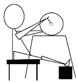
 Subjects rested in the prone position for more than 30 minutes before the experiment. After the H-reflex was elicited at rest, subjects performed arm crank exercise (881E, MONARK) at 60% peakO2 for 10 minutes (60 rpm). Figure 1 shows the scenery during the arm crank exercise. Subjects were not allowed any movements of the lower extremities. Activities of the MG were monitored by recording EMG using an oscilloscope (VC-11, NIHON KOHDEN) during the exercise. When the exercise was completed, subjects changed to prone position keeping the knee joints stable. Then, the H-reflex was measured immediately after, 7, 14, 21, 28, 35, and 42 minutes after the exercise. The H-reflex was measured for less than 2 minutes each time. Subjects maintained the prone position to minimize the variation of the H-reflex caused by changes in position or muscle activity9). The subject's arms were supported by a table in a comfortable position alongside the subject's head. During data collection, the foot of the tested lower extremity rested keeping the ankle angle free. Subjects were not allowed to move and sleep during the measurement. The electrodes were placed by using surgical tape in order to avoid the changes in the position of stimulation of the tibialis nerve and to prevent an influence on the latent time of M wave. Subjects were instructed to avoid intense exercises before the experiment. The temperature and the humidity during the experiment were 25.1±2.4℃ and 36.8±12.6%, respectively.
 Measurement item
 The measurement items were the heart rate (HR) and the latent time of H-reflex. The surface electromyogram (EMG) was recorded in the MG of the right leg. The skin hair was shaved, and the skin was cleaned using alcohol swabs. The inter-electrodes distance was 20 mm. The electrical resistance was less than 10 KΩ, and the time constant was 0.003 sec. The H-reflex of MG was evoked by electrical stimulation (SEN-3301, NIHON KOHDEN) at the tibialis nerve in the popliteal fossa continuously, once per 4-second interval. The electrical stimulation was applied 10 times during H-reflex measurement. The stimulus to elicit the H-reflex was delivered as 1-ms square pulse. EMG data was recorded on a personal computer (Power book G4, Mac) via analog-to-digital converter (PowerLab/800, AD instruments) at 10 kHz.
 Methodology of analysis
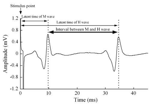
 We compared the interval between the latent time of M wave and H wave, because there was no change in the latent time of M wave each time (Figure 2). The data were expressed as the changes vs. the pre exercise resting level.
 Statistical analysis
 Changes of the HR and the interval between the latent time of M and H wave following the time course were statistically analyzed using one-way analysis of variance (ANOVA). Post hoc comparisons were performed using the Bonferroni test. Statistical analysis was performed using SPSS version 14.0; results were reported as mean±SD. The accepted level of significance was set at 5%.

Fig. 1. Scenery of during the arm crank exercise.

Fig. 2. Methodology of analyses.


RESULTS
 The HR immediately after arm crank exercise was 131±15 beats per min (bpm). The HR immediately after exercise was significantly increased compared to the pre-exercise level. The HR returned to the pre-exercise level during the post-exercise examination.
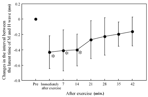
 Figure 3 shows the time course changes in the interval between the latent time of M and H wave. The mean level immediately after exercise was -0.43±0.21 ms as compared to pre-exercise (0 ms). The levels immediately, 7, and 14 min after exercise were significantly shorter than that of the pre-exercise level (P<0.05). However, the levels gradually returned to the pre-exercise level (ANOVA: P<0.05 vs. pre-exercise).
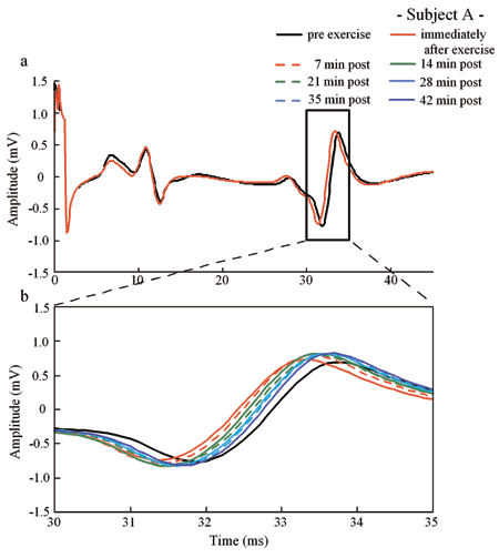
 Figure 4 represents the typical results in subject A, showing the comparison of H-reflex in pre- and post-exercise. Figure 4-a shows the patterns of H-reflex obtained before and immediately after exercise. The latent time of M wave did not change in response to exercise. The interval between the latent time of M and H wave after exercise, however, was shorter than that of the pre-exercise level. Figure 4-b shows the patterns of H-reflex between 30 and 35 ms. The interval between the latent time of M and H wave gradually returned to the pre-exercise level following recovery.

Fig. 3.  Time course changes (Δ) in the interval between the latent time of M and H wave vs. pre-exercise (pre). Mean±SD.
*: P<0.05 vs. pre exercise level.

Fig. 4.  Comparison of the H-reflex in Subject A between pre- and post-exercise. a: pre vs. immediately after exercise. b: recovery of the latent time of H wave. The latent time of M wave did not change in response to arm crank exercise. However, that of H wave immediately after exercise was shortened significantly


DISCUSSION
 Immediately after exercise
 H-reflex showed that the recruitment of the MU of fast-twitch muscle systems was caused by the facilitatory effects of a spinal MN pool by the muscle contractions26). The mean spike amplitude of the recorded MU activities increased as the force level increased according to the analyses of the intramuscular spike amplitude and frequency19). As the recruitment threshold of MUs in isometric conditions follows the size principle, the increased mean spike amplitude would show the activation of newly recruited MUs5,6,12). We considered that the MU of fast muscle system was recruited remarkably immediately after arm crank exercise. The interval between the latent time of M and H wave was shortened. The HR immediately after arm crank exercise was approximately 130 bpm. It has been reported that cardiac sympathetic outflow does not increase during light to moderate dynamic exercise and that the HR response appears to be predominantly adjusted by withdrawal of cardiac parasympathetic nerve activity22,23). On the other hand, sympathetic drive to the heart is considerably augmented during supine bicycle exercise at 50% of maximum voluntary exercise capacity8). It was considered that the exercise at moderate intensity causes the withdrawal of cardiac parasympathetic nerve system activity and outflow of sympathetic nerve system.
 Both warming and cooling induced a significant change in the maximum H-reflex (Hmax) onset latency, especially under warming conditions in which subjects presented shorter reflex onset latencies4). As described previously, the changes in the Na+ voltage-gated channel function affect the conduction of the depolarization24). Hodgkin and Katz11) described a change in temperature altered the opening and closing time of the Na+ channels. Cooling slows down the depolarization time. Na+ ions move into the cell during the prolonged opening, a larger depolarization occurs and the MU action potentials increase, while warming has the opposite effect24). We checked the responses of rectal temperature to the same experimental protocol in 10 subjects after the experiment. The rectal temperature immediately after exercise was significantly elevated by 0.2±0.1℃ as compared to the pre-exercise level. Thus, it is speculated that an additional mechanism to explain the shortened interval between the latent time of M and H wave immediately after exercise may be related to the altered autonomic nerves system and the increased body temperature caused by arm crank exercise.
 Recovery time
 The interval between the latent time of M and H wave, which was shortened immediately after exercise, returned to the pre-exercise level gradually (Figure 3). Immediately after exercise, HR recovery is mainly attributed to vagal reaction1). The rectal temperature begun to decrease approximately 7 min later from the level seen immediately after exercise. These results suggest that the changes of autonomic nervous system activity and body temperature could affect the levels of the recruitment threshold of MU.
 Experiment consideration
 The arm crank exercise was performed in this study and the H-reflex was evoked from the MG, because the position of the electrode for stimulation placed at the tibial nerve did not change and fatigue of the MG did not evoke. It was reported that an afferent impulse from the upper limb influenced the excitability of MNs of the lower limbs10,17). And, human subjects were tested whether the recurrent inhibition of soleus and wrist flexor MNs could be modified by transcranial magnetic stimulation17). It was concluded that the H-reflex was facilitated by Renshaw cells of spinal cord via a short interneuronal chain in both the upper and lower limb17). Therefore, we speculated that the gastrocnemius MN may be facilitated by performing arm crank exercise.
 Research perspective
 This study suggested that arm crank exercise had an influence on the activity of the spinal cord α-MNs. However, the detailed mechanism is still unclear. It will be necessary to examine the influence of autonomic nervous system activity on the spinal cord α-MNs in the future. Moreover, it will be importance to study the connection between temperature and spinal cord α-MNs.
 In conclusion, the interval between the latent time of M wave and H wave immediately after exercise was shortened significantly as compared to that of pre-exercise level. But the interval returned to the pre-exercise level as time progressed during the recovery time.
 
ACKNOWLEDGEMENTS
 The authors gratefully acknowledge Dr. Hiroaki Takekura (Department of Physiological Sciences, National Institute of Fitness and Sports, Kanoya, Kagoshima) for advice on this research. We are grateful to Mr. Michael J. Kremenik and Mr. Tsukasa Toubaru (Department of Health and Sports Science, Kawasaki University of Medical Welfare, Kurashiki) for their generous help in writing of the English manuscript.
 
REFERENCES

 
1) Arai, Y., Saul, J.P., Albrecht, P., Hartley, L.H., Lilly, L.S., Cohen, R.J. and Colucci, W.S.: Modulation of cardiac autonomic activity during and immediately after exercise. Am. J. Physiol., 256, 132-141, 1989.
2) Bigland-Ritchie, B., Cafarelli, E. and Vollestad, N.K.: Fatigue of submaximal static contractions. Acta Physiol. Scand., 128, 137-148, 1986.
3) deVries, H.A., Wiswell, R.A., Bulbulian, R. and Moritani, T.: “Tranquilizer effect of exercise, Acute effects of moderate aerobic exercise on spinal reflex activation level”. Am. J. Phys. Med., 60, 57-66, 1981.
4) Dewhurst, S., Riches, P.E., Nimmo, M.A. and De Vito, G.: Temperature dependence of soleus H-reflex and M wave in young and older women. Eur. J. Appl. Physiol., 94, 491-499, 2005.
5) Fallentin, N., Jorgensen, K. and Simonsen, EB.: Motor unit recruitment during prolonged isometric contractions. Eur. J. Appl. Physiol. Occup. Physiol., 67, 335-341, 1993.
6) Feiereisen, P., Duchateau, J. and Hainaut, K.: MU recruitment order during voluntary and electrically induced contractions in the tibialis anterior. Exp. Brain Res., 114, 117-123, 1997.
7) Fuglevand, A.J., Zackowski, K.M., Huey, K.A. and Enoka, R.M.: Impairment of neuromuscular propagation during human fatiguing contractions at submaximal forces. J. Physiol., 460, 549-572, 1993.
8) Hasking, G.J., Esler, M.D., Jennings, G.L., Dewar, E. and Lambert, G.: Norepinephrine spillover to plasma during steady-state supine bicycle exercise. Comparison of patients with congestive heart failure and normal subjects. Circulation, 78, 516-521, 1988.
9) Hayes, K.C. and Sullivan, J.: Tonic neck reflex influence on tendon and Hoffmann reflexes in man. Electromyogr. Clin. Neurophysiol., 16, 251-61, 1976.
10) Hiraoka, K. and Nagata, A.: Modulation of the soleus H reflex with different velocities of passive movement of the arm. Electromyogr. Clin. Neurophysiol., 39, 21-26, 1999.
11) Hodgkin, A.L. and Katz, B.: The effect of temperature on the electrical activity of the giant axon of the squid. J. Physiol., 109, 240-249, 1949.
12) Houtman, C.J., Stegeman, D.F., Van Dijk, J.P. and Zwarts, MJ.: Changes in muscle fiber conduction velocity indicate recruitment of distinct motor unit populations. J. Appl. Physiol., 95, 1045-1054, 2003.
13) Kurata, H.: Long period constancy of threshold force of signal human motor units in voluntaryy contraction and its change induced by desire for micturtion. Jikeikai Med. J., 23, 103-111, 1974.
14) Löscher, W.N, Cresswell, A.G. and Thorstensson, A.: Excitatory drive to the α-motoneuron pool during a fatiguing submaximal contraction in man. J. Physiol., 491(1), 271-280, 1996.
15) Macefield, V.G. and Elam, M.: Comparison of the firing patterns of human postganglionic sympathetic neurones and spinal alpha motoneurones during brief bursts. Exp. Physiol., 89(1), 82-88, 2004.
16) Mao, C.C., P. Ashby, M.W. and Mccrea, D.: Synaptic connections from large muscle afferents to the motoneurons of various leg muscles in man. Exp. Brain Res., 56, 341-350, 1984.
17) Mazzocchio, A., Rossi, A. and Rothwell, J.C.: Depression of Renshaw recurrent inhibition by activation of corticospinal fibres in human upper and lower limb. J. Physiol., 481, 487-498, 1994.
18) Misiazek, J.E., Brooke, J.D., Lafferty, K.B., Cheng, J. and Staines, W.R.: Long-lasting inhibition of the human soleus H-reflex pathway after passive. Brain Res., 677(1), 69-81, 1995.
19) Moritani, T. and Muro, M.: Motor unit activity and surface electromyogram power spectrum during increasing force of contraction. Eur. J. Appl. Physiol. Occup. Physiol., 56, 260-265, 1987.
20) Moritani, T., Muro, M. and Nagata, A.: Intramuscular and surface electromyogram changes during muscle fatigue. J. Appl. Physiol., 60(4), 1179-1185, 1986.
21) Onodera, S., Morimoto, S. and Kasuga, N.: Relation Between the Functional of the Autonomic Nervous System and Voluntary Movement. Descente Sports Sci., 4, 265-270, 1983.
22) Robinson, B.F., Epstein, S.E., Beiser, G.D. and Braunwald, E.: Control of heart rate by the autonomic nervous system. Studies in man on the interrelation between baroreceptor mechanisms and exercise. Circ. Res., 19, 400-411, 1966.
23) Rowell, L.B. and O'Leary, D.S.: Reflex control of the circulation during exercise: chemoreflexes and mechanoreflexes. J. Appl. Physiol., 69, 407-418, 1990.
24) RutKove, S.B.: Effects of temperature on neuromuscular electrophysiology. Muscle Nerve, 24, 867-882, 2001.
25) Sakamoto, K. and Swie, Y.W.: EMG characteristics of low back and lower limb muscles during forward bending posture. Elecromyogr. Clin. Neurophysiol., 43, 335-347, 2003.
26) Tanaka, S. and Nagata, A.: Motoneuron Pool on The Spinal Cord by The Measure of H wave Spectrum. A new development of time series analysis in medical and biological sciences., Hokkaido University Press, Hokkaido, pp. 703-716, 1996.
27) Trimble, M.H. and Harp, S.S.: Postexercise potentiation of the H-reflex in humans. Med. Sci. Sports Exerc., 30(6), 933-941, 1998.


Send correspondence to:
     Doctoral Program in Health Science, Graduate School of Kawasaki University of Medical Welfare, 288 Matsushima, Kurashiki, Okayama, 701-0193 Japan
     Kazutoshi SEKI
     TEL: 81-86-462-1111(54531)
     FAX: 81-86-464-1109
     E-mail:w6304005@mw.kawasaki-m.ac.jp